Capnocytophaga canimorsus

capnocytophaga canimorsus

El Capnocytophaga canimorsus es una bacteria Gram negativa que causa una enfermedad zoonótica, especialmente en pacientes sin bazo. La bacteria es parte de la flora normal de las encías de perros y gatos. La enfermedad causada por C. canimorsus cursa con sepsis potencialmente fulminante por asociación a coagulación intravascular diseminada.

capnocytophaga canimorsus 2

1.- Un varón de 50 años, esplenectomizado por una PTI, presenta, tras ser mordido por un perro, un cuadro de  fiebre, hipotensión, malestar general, leucocitosis, trombopenia y aumento de los tiempos de coagulación. Respecto a la entidad que presenta este paciente, ¿cuál es el tratamiento de elección?

1- Doxiciclina.
2- Ciprofloxacino.
3- Ceftazidima.
4- Rifampcina.
5- Penicilina.

R.C: 5

CAPNOCYTOPHAGA CANIMORSUS 1


2.-¿Cuál es el gérmen más frecuentemente implicado en los cuadros de sepsis fulminante con shock, CID y meningitis en pacientes alcohólicos /cirróticos que han sido mordidos por animales?

1- P. aeuriginosa

2- Afipia felis

3-Capnocytophaga canimorsus

4-Eikenella corrodens

5- Streptobacilus moniliformis

R.C: 3

Apical hyperthrophic cardiomyopathy


Apical

Apical hypertrophic cardiomyopathy (AHC) is a rare variant of hypertrophic cardiomyopathy. It was described by Sakamoto in 1976 in Japanese patients.  Here there is hypertrophy only in apex. ECG tipically shows  giant negative T  waves in anterior precordial derivations. Ventriculography or magnetic resonance is tipically too: we see the ventricule like ¨AS OF PICAS¨.

Slide 1


apicalcardiomiopatia as picas


Four-chamber steady-state free precession cine MR image makes the apical endocardial border (arrows) clear. Two-chamber steady-state free precession cine MR image shows apical hypertrophy (arrows) and obliteration of the LV apical cavity at end diastole, with a typical spadelike configuration.


MIOCARDIOPATÍA HIPERTRÓFICA APICAL (SEPTO BAJO).

Es una variante rara de la miocardiopatía hipertrófica. Fue descrita por Sakamoto en 1976 en pacientes japoneses. En esta miocardiopatía existe hipertrofia sólo a nivel del ápex. Se caracteriza por presentar ondas T gigantes negativas en el ECG en las derivaciones precordiales. La ventriculografía o la resonancia magnética son muy típicas también, en las que la cavidad ventricular izquierda adopta forma de naipe o ¨AS DE PICAS¨. Suele tener un curso clínico benigno.

En las imágenes de resonancia magnética se puede observar una clara hipertrofia y obstrucción del ventrículo izquierdo apical al final de la diástole con la típica configuración de naipe.

Paroxysmal nocturnal hemoglobinuria (PNH)


¨A 32-year-old woman presented to the emergency department with a 3-day history of epigastric pain, vomiting, increasing pallor, and dark urine. Her medical history disclosed no abnormal findings. Physical examination revealed mild jaundice, abdominal distention, and bilateral lower extremity edema. Laboratory studies disclosed anemia, with a hemoglobin level of 70 g/L (7.0 g/dL), where the normal range is 115–160 g/L (11.5–16.0 g/dL). Hyperbilirubinemia was also disclosed, with a total serum bilirubin level of 75.2 μmol/L (4.4 mg/dL), where the normal range is 0–17.1 μmol/L (0–1.0 mg/dL). Liver enzyme levels were elevated, with an alanine aminotransferase, or ALT, level of 51 U/L (normal range, 5–37 U/L) and an aspartate aminotransferase, or AST, level of 170 U/L (normal range, 10–37 U/L). Serum creatinine level was normal. Findings at abdominal radiography showed a small amount of bowel gas but disclosed no other abnormalities. Abdominal ultrasonography (US) , contrast material–enhanced computed tomography (CT) , and contrast-enhanced magnetic resonance (MR) imaging  were performed, in that order.¨

IMAGING FINDINGS

US depicted an echogenic thrombus in the intrahepatic inferior vena cava, a prominent caudate lobe, and mild diffuse heterogeneity of the liver parenchyma. Contrast-enhanced CT  depicted a filling defect in the inferior vena cava that extended into the left hepatic vein, preferential enhancement of the central liver and an enlarged caudate lobe, mild ascites, and a filling defect in the portal vein. Findings at contrast-enhanced MR imaging  confirmed the filling defects in the inferior vena cava and portal vein and also showed a diffuse reduction in renal cortical signal intensity.

Marchiafava-Micheli syndrome

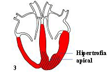
It is corpuscular ADQUIRED hemolytic anemia due to  the mutation of gen PIG-A in hematopoietic cells . All of hemolytic anemia are congenital except this type of corpuscular anemia. All of our cells have two molecules in the membrane called CD55 and CD59. They protect us from the action of the complement which produces their lysis. If we don´t have these molecules, the complement lysis of cells and we will have hemolytic anemia, leucopenia and trombopenia (PANCITOPENIA) and red urine.  But be careful, although we see trombopenia , patiens suffer trombosis in rare localizations like suprahepatic veins (Budd- Chiari Syndrome) and that´s the reason why this patients could die.

HPN

DIAGNOSIS

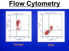
Flow cytometry (we will not see CD 55 or CD59).

Flow Citometry

Ham test:  The test involves placing red blood cells in mild acid. The acid induces the activation of the complement and lysis of red blood cells.

Sucrose test: induces too the activation of the complement and hemolysis.

TREATMENT

1) Steroids

2) Anticoagulation

3) Blood transfusion

4) Fe

5) ECULIZUMAB: anti C5 complement

ECULIZUMAB

6) Hematopoietic stem cell transplantation: curative treatment

http://radiology.rsna.org/content/225/1/67.full

The ¨Broken Heart Syndrome¨: Takotsubo Cardiomyopathy

Case Report: Chest pain after emotional and physical upset

A 61 year old white woman attended her local accident and emergency department with severe central chest pain. After being chased by two large dogs. The pain was not relieved by nitroglycerine spray given in the ambulance. An electrocardiogram showed anterolateral ST segment depression, with an elevated troponin T of 1.25 μg/l. She had no cardiovascular risk factors. Non-ST elevation myocardial infarction was diagnosed, and the patient was treated accordingly.
Coronary angiography showed normal coronary arteries, but the left ventriculogram showed a large area of apical hypokinesis with moderate impairment of left ventricular systolic function.
The patient was readmitted several weeks later with further chest pain. An electrocardiogram showed no new changes with no rise in the cardiac troponin. An echocardiogram showed that her left ventricular systolic function had almost returned to normal.
broken heart
blog_entry_148290
Takotsubo