Peptic ulcer.Úlcera péptica

ASKANAZY´s levels.Capas de ASKANAZY. 

Regla mnemotécnica/ mnemonic: NIGgaS
1) N– necrosis. Restos fibrino- Necróticos superficiales.

2) I– inflamation.  Infiltrado inflamatorio con numerosos neutrófilos (II PMN-N)

3) G– Granulosis. Tejido de granulación jóven.

4) FibrosiS. Tejido fibroso cicatricial.

askanazy

ENFERMEDAD DE PAGET DE LA MAMA

Se trata de una enfermedad de la piel en la que hay una infiltración de la dermis de la areola y el pezón por un carcinoma mamario de crecimiento intracanalicular. Histológicamente se caracteriza por la presencia de células de Paget en la epidermis. Son células grandes, de citoplasma eosinófilo y PAS+. Clínicamente se presenta como un eczema a nivel del pezón /areola que no cura con el tratamiento corticoideo.

paget disease 1 Paget disease

TOF

Chest x-ray findings which are considered characteristic for various congenital heart diseases:

A)Boot-shaped heart: Tetralogy of Fallot, tricuspid atresia
B)Egg-shaped heart: Transposition of great arteries
C)Snowman silhouette: Total anomalous pulmonary venous return (supracardiac)
D)Rib notching: Coarctation of the aorta (older children)

tof

Case of CVS dis


Which one of the following conditions is included in the differential diagnosis for high-output heart failure?

A. Biotin deficiency.
B. Hyperparathyroidism.
C. Paget’s disease.
D. Pulmonary hypertension.

In the latest Clinical Problem-Solving article, a 63-year-old woman presented with edema and erythema of the skin of her legs and with orthopnea and paroxysmal nocturnal dyspnea. The edema had first appeared almost 2 years earlier but had worsened markedly in the previous week.

Isolated, bilateral lower-extremity swelling and erythema that have been stable over a long period suggest venous stasis, but an abrupt worsening of edema along with symptoms of orthopnea and paroxysmal nocturnal dyspnea warrants prompt evaluation for other possible causes of such swelling, including cardiac, hepatic, and renal dysfunction. Lymphedema can cause profound lower-extremity edema but should not cause orthopnea or exertional dyspnea.

Clinical Pearls

• What is in the differential diagnosis of high-output heart failure?

The differential diagnosis for high-output heart failure includes thiamine deficiency, hyperthyroidism, severe anemia, Paget’s disease, and arteriovenous fistula.

• What are the clinical characteristics of high-output heart failure?
High cardiac output is defined as a cardiac output greater than 8.0 liters per minute (or a cardiac index >4.0 liters per minute per square meter). The initial physiological derangement is usually low systemic vascular resistance, which incites a neurohormonal response culminating in the retention of salt and water and in volume overload. Patients with high-output heart failure often have signs similar to those seen in a low-output state (pulmonary congestion, peripheral edema, and hypotension), but the presence of a wide pulse pressure (in the absence of clinically significant aortic regurgitation) and warm, well-perfused extremities can help differentiate the two entities.

Morning Report Questions

Q: How is high-output heart failure diagnosed?

A: Echocardiography is a sensitive tool for identifying the elevated cardiac filling pressures, enlarged cardiac chambers, and supranormal stroke volume that accompany a high-output state. A high velocity-time integral on echocardiography may be an early clue to the presence of a high-output state. Cardiac catheterization, with hemodynamic assessments and a shunt study, is usually required to make a definitive diagnosis.

Q: What are the cardiovascular characteristics of thiamine deficiency?

A: High cardiac output can be driven by nutritional deficiencies (e.g., with wet beriberi caused by thiamine deficiency). In thiamine deficiency, the severity of cardiomyopathy does not correlate reliably with laboratory thiamine assays. Measurement of erythrocyte thiamine transketolase activity (with and without thiamine loading) is a more specific assessment for thiamine deficiency than is measurement of the serum thiamine level and is the preferred confirmatory test, though the whole-blood assay is a validated alternative. Most cases of thiamine deficiency are attributable to malnutrition, but this deficiency can also develop as a result of long-term administration of diuretics in patients who are being treated for heart failure.

CORAZON

BOTULISMO

botulism

1) DISFUNCIÓN BULBAR: disartria, disfagia

2) FALLO PARASIMPÁTICO:

botulismo miel

  • Sequedad de ojos y boca
  • íleo paralítico
  • Estreñimiento(inicialmente puede aparecer diarrea por irritación intestinal)
  • Retención urinaria
  • MIDRIASIS

3) PARÁLISIS DESCENDENTE SIMÉTRICA

  • VI: 1º en afectarse
  • III: diplopia, ptosis, midriasis
  • Disartria, disfagia ,después se afectan BRAZOS y PIERNAS y FINALMENTE LA MUSCULATURA RESPIRATORIA (MUERTE).

botulismtoxinaction

1) A 2-mo-old infant is suspected of having infant botulism. Which is the best means to confirm the diagnosis? QUESTIONS:

A-Lumbar punctureimages
B-CT scan
C-Muscle biopsy
D-Electromyography
E-Fecal specimen

E-Fecal specimen

Explanation: The diagnosis of botulism is unequivocally established by demonstrating the presence of botulinum toxin in serum or of C. botulinum toxin or organisms in wound material or feces.

2)Which of the following findings on the history or physical examination excludes a diagnosis of infant botulism?

A-No ingestion of honey or corn syrup
B-Normal latch, suck, and swallow
C-Soft feces
D-Normal deep tendon reflexes
E-Metabolic acidosis

B-Normal latch, suck, and swallow
Explanation: In infants, bulbar palsies are evident as poor feeding, weak suck, feeble cry, drooling, and even obstructive apnea


4d40143f7e0c6

Anthrax

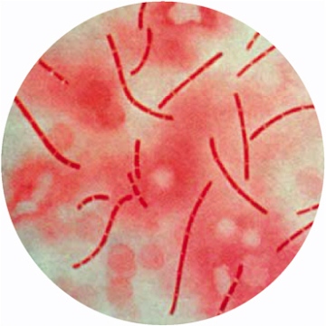
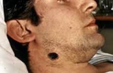
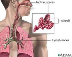
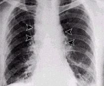
Imagen Infectious disease caused by Baccilus anthracis, a gram positive aerobic bacterium. It is transmited by ingestion (intestines), inhalation (lungs), or cutaneous (skin). Commonly animals herbivors  (bovines) are infected and they transmited the disease to human. The skin form has a  very cleary sign, there is a point which is ulcerative and with a blue colour, reason why in some countries this disease is called BLUE SPOT.   The intestinal form is more rare and it seem gastroenteritis . The lungs could be afected too, the clinical sintoms are inespecific, but this form of the disease is very severy. We must make  X-ray of the lungs (there is an ensachement of the mediastine). In the anamnesis is important to ask for the proffesion of the patient.  The confirmation of our diagnosis is made by demostration of the bacteria in blood, feces, expectorations or skin lesions. The treatment is based on fluorquinolones like ciprofloxacine, doxicicline in combination with penicilin. The corticosteroids are useful for the treatment of the inflamation and oedema .

ImagenImagen

Imagen

Imagen


ImagenImagen